3D and Mixed Reality Transform Bone Cancer Surgery
An Arthroplasty article details how mixed reality and custom-made guides achieve more precise, bone-preserving osteosarcoma resections than freehand techniques. The Yale School of Medicine study shows the potential of these technologies to reduce resection failure while protecting joints and bone.
The operation for osteosarcoma aims to remove the tumour in its entirety. Surgical strategy has long relied on a wide margin approach, in which healthy bone surrounding the lesion is excised to create a buffer zone. Such margins are intended to reduce the risk of residual malignant cells remaining after surgery. The wide margin technique, however, can result in considerable loss of healthy tissue, sometimes necessitating joint sacrifice, limb shortening or, in extreme cases, amputation. Conversely, overly conservative cuts risk leaving viable tumour cells behind.
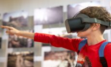
For years, surgeons have removed tumours using freehand methods, mentally translating 2D scans to bone markings. Limited visibility and access can make accurate cuts difficult.
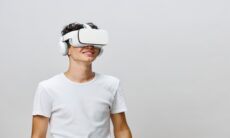
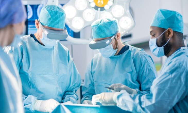
The study in Arthroplasty examined two technology-assisted alternatives. The first used 3D printing to produce patient-specific cutting guides that fit a single bone surface. These guides include slots that control the saw blade’s path, thereby constraining the cut to a preplanned trajectory. The second used mixed-reality headsets to superimpose a holographic surgical plan over the patient’s anatomy, aligning a digital blueprint with the physical bone in the operating field.
Lab testing on anatomical models showed clear differences between methods. Freehand cuts varied more in placement and angle, sometimes too close to the tumour or excessively wide. The 3D-printed guides and mixed reality yielded narrower, consistent margins, allowing cuts closer to malignant tissue with no clear increase in risk.
Physical guides provided mechanical stability through their fit and blade slots. Mixed reality provided visual adjustability and a 360-degree view without the need for plastic guides. The holographic overlay helped the team see the tumour’s extent during surgery, improving spatial cognition.
Patient-specific instruments also help with planning. Engineers create exact bone replicas before surgery, allowing them to simulate cuts and consider bone density and saw blade kerf. This makes it easier to design guides that account for kerf loss, which is hard to do by sight alone.
The authors note that technology can’t replace surgical skill. They saw digital model instability, showing the risk of technical failure. If mixed reality fails, surgeons must switch to manual methods. Tactile feedback, like feeling bone and tool vibration, remains vital; visual guides support but do not replace hands-on skill.
The study notes ongoing work to improve robustness. Researchers are developing methods to ensure holograms remain fixed to the bone despite bone movement or alterations in the surgeon’s viewpoint. Further clinical research is required to establish how laboratory results translate into clinical practice and to determine comparative outcomes across larger patient cohorts.
The research supports more conservative, function-preserving limb-salvage surgery using 3D and mixed reality, signalling steady progress toward better patient results and quality of life.
The paper lists the principal investigator and co-authors as follows: Steven Tommasini, Jose Caceres-Alban, Dieter Lindskog, Johannes Sieberer, and Alyssa Glennon. The research was carried out as part of a master’s programme in personalised medicine and applied engineering in collaboration with 3D Collaborative for Medical Innovation.